With the remarkable potential of isotopic analysis making recent headlines (see p.18), it seems apt to talk a bit more about this technique. Among the wealth of archaeological questions isotope analysis can help to answer are: where was an individual born and raised, did they migrate during their lifetimes, what did they predominately eat, and when were they weaned? As this is a relatively new and ever-evolving methodology, though, some of the wrinkles are still being ironed out – and one of the biggest questions currently being explored is whether bone is as effective as teeth in reflecting the isotopic values that a person accumulated in life.
One of the problems with bone is that it is relatively porous, allowing the isotopic signature of the burial environment to contaminate any remains buried within it (although, as we see in this month’s Stonehenge feature, cremated bone may not be as susceptible to such pollution). Tooth enamel, on the other hand, has a much tighter structure, which makes it relatively impervious and hence more accurately represents an individual’s isotopic signature during life.
But the burial environment may not be the only factor affecting the accuracy of isotope values in bone. A new study based on human remains from the Anglo-Saxon cemetery of Raunds Furnells (c.AD 978-1040; see CA 106) in eastern Northamptonshire has shown that, during childhood, bone growth responds differently to periods of biological stress than tooth formation does, which has an impact on the isotopic levels that it incorporates. Taking a single tooth from 18 children and five adults buried at the site, the team – from the universities of Bradford, Sheffield, Bournemouth, Aberdeen, and Durham – carried out carbon and nitrogen isotopic analyses on each individual, and then compared it with already published data on the isotope ratios present in the bones of those same individuals.
In order to assess how these ratios changed over an individual’s first few years of life, they took thin, horizontal sections of each tooth, all the way from the crown to the root. They could thus assess any changes in the signature between when the tooth began to form and once it was fully developed. They were able to do this because teeth form incrementally: they start at the crown and finish forming at the root. The first 0.5mm of our milk teeth’s crowns are formed when we are still foetuses. After we are born, more and more of the tooth is formed, until around 2.5 years when the last bit of the root is laid down – this process is the same for all teeth, but the timing varies depending on the tooth. If we take horizontal slices of these teeth each section of enamel provides a unique isotopic signature that will reflect the isotopes that individual was consuming during the stage of their life when that part of the tooth was formed. So, for milk teeth, a sample from the top of the crown will give us information about an individual in utero, while a sample from the bottom of the root will tell us about their life from around 2.5 years of age.
The team was primarily interested in ascertaining how accurate isotopic analysis is in assessing weaning. This is usually determined by detecting shifts in the isotopic ratios between infants and women of child-bearing age. During pregnancy, the isotopic values of the mother and child should be similar, but breastfeeding results in a rise in these values. They then start to fall during the weaning process, once other foods are introduced. This characteristic curve allowed the team to detect different patterns of breastfeeding and weaning in the individual tooth samples. But, as the team discovered, the nitrogen and carbon isotope values measured in the bone and teeth samples from the 23 Raunds Furnells individuals were not the same. On average, the bone data were recording lower nitrogen isotope values than teeth forming at the same time, meaning that the two tissues were indicating different weaning patterns within the same individual.
In the paper presenting the results of this project, recently published in the American Journal of Physical Anthropology (https://doi.org/10.1002/ajpa.23682), the team suggests that this discrepancy may be caused by the fact that stress – whether nutritional, physiological, or emotional – is more likely to disrupt the growth of bone than it is the formation of dentine. When bone growth stops, it is not taking in any more isotopes, and hence does not ‘record’ the person’s signature during the period of time when growth is arrested. This appears to be particularly true the younger the individual is: for all the individuals that survived past the age of three, their bone isotopic values matched those of the teeth.
These results show that bone can provide a very inconsistent isotopic profile, particularly if the individuals being assessed have undergone significant periods of stress – as many of the individuals from the Raunds Furnells cemetery appear to have done. This suggests that in future, teeth may be the preferred sample to use in isotopic weaning studies.
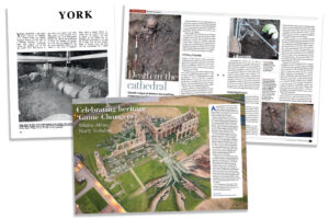
This article appeared in CA 344.